Introduction
Legalization of cannabis in various US states and other countries lead to a development of efficient analytical tool for law-enforcement officers to evaluate D9-tetrahydrocannabinol (THC) (major active metabolite of Cannabis). When consumption ends, THC levels in blood decrease rapidly, giving a short period of time for police enforcement to collect blood.
For this project, we propose to perform a liquid-liquid extraction method for THC in blood collected with a Mitra device. Quantification using the Luxon coupled to a mass spectrometer (Luxon-MS/MS) is chosen as a fast-analytical technique.
Luxon Ionization Source
The Luxon Ion Source® (Figure 1) is the second-generation sample introduction and ionization source based on the LDTD® technology for mass spectrometry. Luxon Ion Source® uses Fiber-Coupled Laser Diode (Figure 2) to obtain unmatchable thermal uniformity giving more precision, accuracy and speed. The process begins with dry samples which are rapidly evaporated using indirect heat. The thermally desorbed neutral molecules are carried into a corona discharge region. High efficiency protonation and strong resistance to ionic suppression characterize this type of ionization and is the result of the absence of solvent and mobile phase. This thermal desorption process yields high intensity molecular ion signal in less than 1 second sample-to-sample and allows working with very small volumes.
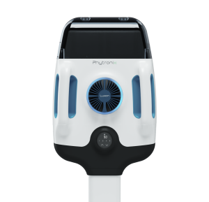
Figure 1 – Luxon Ion Source®
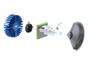
Figure 2 – Schematic of the Luxon Ionization Source
Sample Preparation Method
THC is spiked in human EDTA-k2 blood at concentrations ranging from 2.5 to 100 ng/mL. 10 µL blood samples are then collected with a Mitra micro sampling device (Figure 3).
The blood collector part of the device is transferred in a glass tube (10X75mm) containing 100 µL EDTA buffer (500 µg/mL). Tubes are put in a sonicator bath for 10 minutes. 10 µL of internal standard (THC-d3, 500 ng/mL in acetonitrile) and 100 µL Hexane:Ethyl Acetate (90:10) are added. The solution is then vortexed and centrifuged at 5000 rpm for 2 minutes. Finally, 8 µL of the upper layer are spotted into LazWell™96 plates and evaporated to dryness. Luxon-MS/MS analysis is performed after a complete evaporation.

Figure 3 – Mitra micro sampling device (10 µL)
LDTD-MS/MS Parameters
LDTD
Model: Phytronix, Luxon T-960
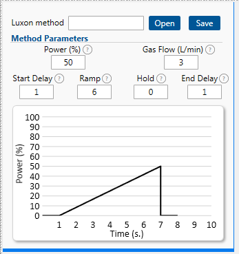
Figure 4 – LUXON parameters
MS/MS
Model: TSQ Altis plus, Thermo Fisher Scientific
Ionization: APCI (negative)
| Molecules | Transition | CE |
|---|---|---|
| THC | 313 → 245 | 30 |
| THC- D 3 | 316 → 248 | 30 |
Results and Discussion
Linearity
A THC calibrator set spiked at the following calibration range are extracted: 2.5 to 100 ng/mL. Correlation coefficients are equal or greater than 0.99 for the quantification curve of THC. Figure 5 shows the calibration curve of THC. Figure 6 shows a typical THC desorption peak with a 0.16-minute window.
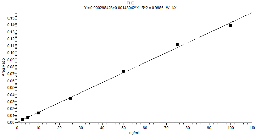
Figure 5 – THC calibration curve
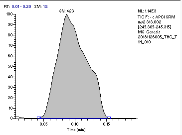
Figure 6 – THC desorption peak
Precision and accuracy
Replicate extractions of control (LLOQ, QC-Low, QC-Medium, QC-High and ULQC) are deposited on a LazWell™ plate and dried before analysis. Replicas are analyzed for inter-run evaluation. CV% below 15% and accuracy within ±15% of nominal concentration are reached. Table 2 shows the inter-run precision/accuracy results.
| THC | LLOQ | QC-Low | QC-Med | QC-High | ULQC |
|---|---|---|---|---|---|
| Conc. (ng/mL) | 2.5 | 5 | 25 | 75 | 100 |
| N | 6 | 6 | 6 | 6 | 6 |
| Mean (ng/mL) | 2.4 | 5.2 | 24.1 | 78.2 | 96.5 |
| %CV | 12.7 | 11.4 | 6.8 | 3.4 | 0.9 |
| %NOM | 95.8 | 104.1 | 96.6 | 104.3 | 96.5 |
THC recovery on Mitra device
Spiked samples of THC (100 ng/mL) are added on a Mitra device (10 µL). The concentration of THC is evaluated against a calibration curve where 10 µL of spiked blood sample are added directly in an extraction tube (100% Mitra recovery). A recovery of 71.5% is obtained. Results are shown in Table 3.
| THC | Recovery |
|---|---|
| Conc. (ng/mL) | 100 |
| N | 6 |
| Mean (ng/mL) | 71.5 |
| %CV | 3.0 |
| %Recovery | 71.5 |
THC stability
THC is a hydrophobic and unstable drug. Different stability parameters are verified. All stability results are shown in Figure 7.
Wet stability in buffer at room temperature and 4°C
After blood collection with the Mitra device, absorbent parts are transferred in glass tubes containing an EDTA buffer. The tubes are kept at room temperature for 1 and 3 hours, then extracted. After 3 hours, in the buffer at room temperature, the THC concentration drops below 80% of the nominal value, but the results after 1 hour are within acceptable nominal values (between 85% to 115%). The same experiments are performed at 4°C for 1, 3 and 20 hours. After 20 hours, in the buffer at 4°C, the THC concentration is still within the acceptable nominal value range.
Wet stability in buffer and extraction solvent at 4°C
After a complete extraction with the Mitra device, the mixture of absorbent parts, EDTA buffer and extraction solvent is kept at 4°C for 1, 3 and 20 hours. After 20 hours, the THC concentration is still within the acceptable nominal value range (between 85% to 115%).
Dry stability of samples on Mitra device
Blood samples are collected with the Mitra device and kept at room temperature for 1, 3 and 20 hours. Then, the absorbent parts are transferred in glass tubes containing the EDTA buffer and an LLE is performed. After 3 hours (blood sample on Mitra device at room temperature), the THC concentration drops below 80% of the nominal value, but the results after 1 hour are within the acceptable nominal value (between 85% to 115%) value.
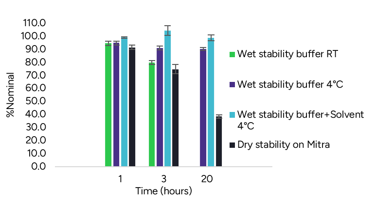
Figure 7 – THC stability results
Conclusion
A low blood sample volume (10 µL) can be properly collected with the Mitra device for THC analysis. THC analysis needs particular care when dealing with the stability of samples. Luxon Ion Source® combined to Thermo TSQ Altis plus mass spectrometer system allows ultra-fast (8 seconds per sample) quantification of THC.